
Posterior Vitreous Detachment
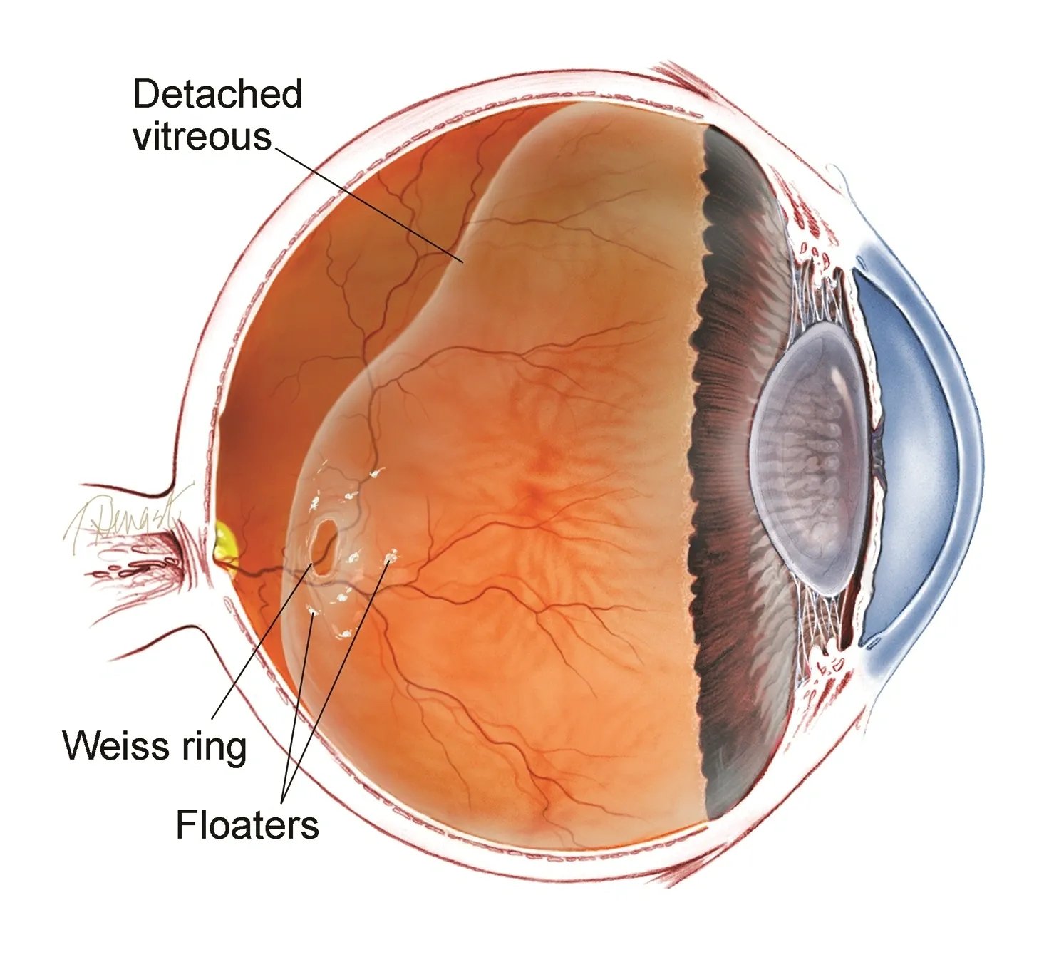
Diagram of Posterior Vitreous Detachment
The vitreous is a clear substance within the eye, which has a gel-like consistency. It fills the hollow space between the lens and the retina at the back of the eye. The vitreous helps maintain the shape of the eye, it absorbs shock and it helps keep the retina in contact with the back wall of the eye.
With ageing the vitreous can become liquified and as a result it can pull away or separate from the retina and collapse into the central hollow space of the eye. This is known as a posterior vitreous detachment (PVD) and it is a normal ageing process.
Risk Factors?
Ageing
Trauma: knock to the head or eye can cause an early onset PVD
Nearsightedness (myopia)
PVD in the other eye
Symptoms?
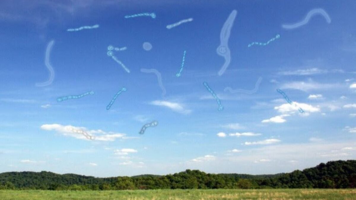
Floaters: These may appear as hundreds of small spots or objects (e.g. cobwebs, squiggly lines) “floating” in the field of vision. These floaters may be black, grey or clear in colour.
Lightning flashes: These may appear as a “sparkle”, “streak” or “twinkle” across the vision.
Decreased vision
The symptoms of a posterior vitreous detachment can often present similar to a retinal tear (or early detachment) or the PVD itself can lead to a retinal tear so it is important to see an ophthalmologist within 24-48 hours of your symptoms presenting to check the health of the retina.
Treatment?
If the posterior vitreous detachment has occurred without associated retinal tears, therapy is not required or indicated. The vitreous will continue to age and liquefy and floaters will usually become less and less noticeable. With time, most people’s symptoms will completely disappear.
Download this information: Posterior vitreous detachment (pdf)

What Eye Floaters look like in a patient with Posterior Vitreous Detachment